Gas exchange in humans
Gas-exchange surfaces
A gas-exchange surface needs:
- a large surface area, providing many sites for diffusion;
- a thin surface, giving a short diffusion distance;
- a good blood supply, maintaining concentration gradients; and
- good ventilation, replacing air and maintaining concentration gradients.
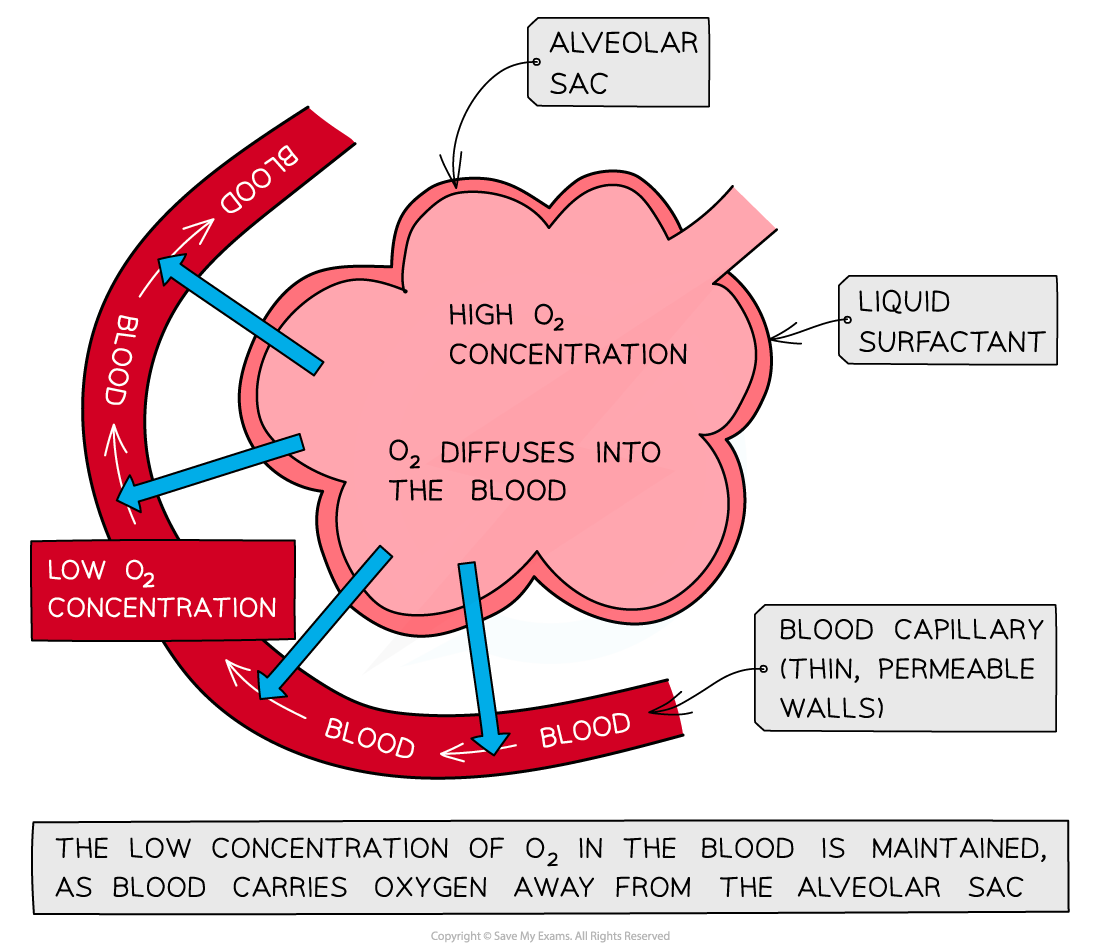
Alveoli provide these features. Their large total surface area gives many diffusion sites, their walls are one cell thick, capillaries carry away oxygen and bring carbon dioxide, and ventilation continually refreshes the air.
Breathing system
| Structure | Function |
|---|---|
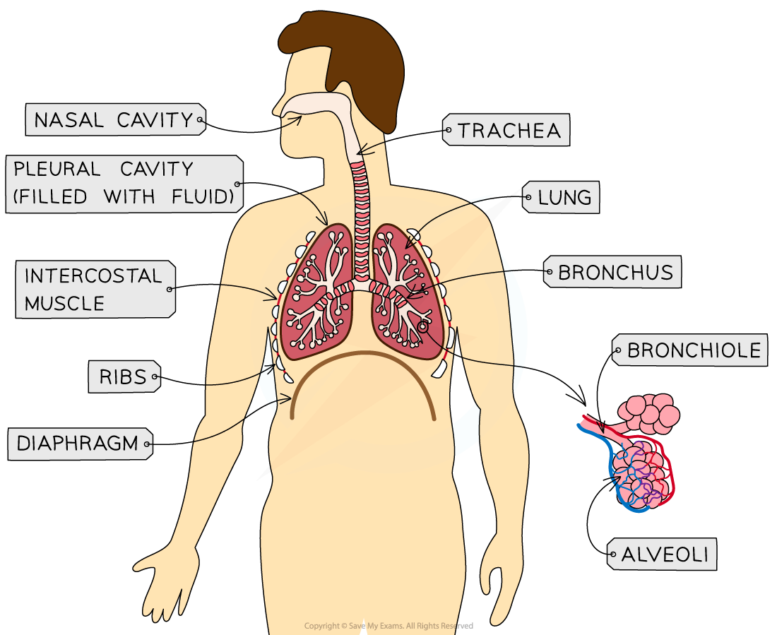
| Lungs | Contain bronchioles, alveoli, and capillaries; main gas-exchange organs. |
| Diaphragm | Muscle that changes thoracic volume. |
| Ribs | Protect the lungs and move during ventilation. |
| Intercostal muscles | Move the ribs during ventilation. |
| Larynx | Connects the throat to the trachea and contains the voice box. |
| Trachea | Carries air towards the bronchi. |
| Bronchi | Two branches carrying air to the lungs. |
| Bronchioles | Narrow branches carrying air to alveoli. |
| Alveoli | Air sacs where oxygen and carbon dioxide are exchanged. |
| Capillaries | Surround alveoli and carry blood for gas exchange. |
Cartilage rings support the trachea and prevent it collapsing while still allowing it to bend.
Ventilation
Ventilation is movement of air into and out of the lungs. Air moves because pressure inside the lungs changes when thoracic volume changes.
Inhalation (inspiration)
- The external intercostal muscles contract, moving the ribs up and out.
- The diaphragm contracts and flattens.
- Thoracic volume increases.
- Pressure inside the lungs decreases below atmospheric pressure.
- Air moves into the lungs.
Exhalation (expiration)
- The external intercostal muscles and diaphragm relax during quiet breathing.
- The ribs move down and in and the diaphragm returns to a dome shape.
- Thoracic volume decreases.
- Pressure inside the lungs increases above atmospheric pressure.
- Air moves out.
During forced expiration, the internal intercostal muscles contract and pull the ribs down more strongly.
Gas exchange at alveoli
Oxygen concentration is higher in alveolar air than in deoxygenated blood, so oxygen diffuses through the alveolar and capillary walls into the blood. It then binds to haemoglobin in red blood cells.
Carbon dioxide concentration is higher in the blood than in alveolar air, so carbon dioxide diffuses into the alveoli and is removed during expiration. Both gases move down concentration gradients.
Inspired and expired air
| Component | Inspired air | Expired air | Reason for change |
|---|---|---|---|
| Oxygen | About 21% | About 16% | Some oxygen diffuses into blood and is used in respiration. |
| Carbon dioxide | About 0.04% | About 4% | Carbon dioxide is produced by respiration and diffuses into alveoli. |
| Water vapour | Less | More, usually close to saturated | Air becomes moist inside the lungs. |
Limewater investigation
Limewater becomes cloudy when it reacts with carbon dioxide to form a calcium carbonate precipitate.
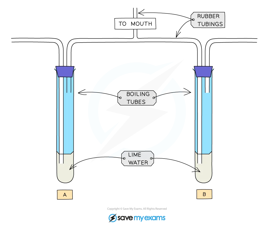
- Place equal volumes of limewater in two identical tubes.
- Pass a sample of inspired air through one tube and expired air through the other, using the same volume or flow rate.
- Compare the time taken for each sample to become cloudy, or compare cloudiness after the same time.
- Repeat measurements and keep limewater volume, apparatus, and air volume constant.
Expired air should turn limewater cloudy faster because it contains more carbon dioxide.
Airway protection
- Goblet cells secrete mucus.
- Mucus traps dust, microorganisms, and other particles and keeps airway surfaces moist.
- Ciliated cells have beating cilia that move mucus towards the throat, where it can be swallowed or expelled.
Physical activity and breathing
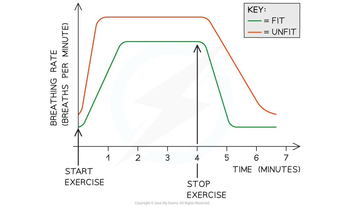
During exercise, muscles respire faster. They require more oxygen and produce more carbon dioxide. Increased carbon dioxide in the blood is detected by the brain, which increases breathing rate and depth. Faster, deeper ventilation supplies more oxygen and removes carbon dioxide.
Investigation
- Allow a subject to rest, then measure breathing rate by counting breaths for a fixed time.
- Measure breathing depth using a suitable method, such as a spirometer, if available.
- Perform a standardised exercise for a fixed time and immediately measure rate and depth again.
- Continue measurements during recovery until values approach the resting level.
- Repeat trials and calculate means.
Keep exercise duration and intensity, subject position, measurement time, and environmental conditions constant. Use the same subject for comparisons where appropriate, and stop if the subject feels unwell. Plot breathing rate and depth against time or exercise condition.
Exam checks
- Ventilation is movement of air; gas exchange is diffusion of oxygen and carbon dioxide.
- External intercostals help inspiration; internal intercostals are important in forced expiration.
- Increased thoracic volume lowers lung pressure; decreased volume raises it.
- Explain exercise responses through increased respiration, carbon dioxide detection, and increased breathing rate and depth.
- In practical questions, name the variable measured, controls, repeats, and expected result.
Introduction
Gas exchange in humans is the process by which oxygen enters the body and carbon dioxide is removed. This occurs primarily in the lungs through a system of specialized structures optimized for efficient gas transfer.
Features of Gas Exchange Surfaces
Four Essential Features
For effective gas exchange, the surface must have four key characteristics:
| Feature | Purpose | How it Works |
|---|---|---|
| Large surface area | More area for gas exchange | Many alveoli create large total area (~70 m² in adult lungs) |
| Thin surface | Short diffusion distance | Alveoli and capillary walls are one cell thick (~0.5 μm) |
| Good blood supply | Maintain concentration gradient | Capillaries carry away O₂, bring CO₂ |
| Good ventilation | Maintain concentration gradient | Breathing brings in O₂, removes CO₂ |
How These Features Work Together
- Large surface area = more sites for O₂ and CO₂ to cross
- Thin surface = faster diffusion (shorter distance)
- Good blood supply = blood constantly removes O₂, loads with CO₂
- Good ventilation = fresh air constantly replaces stale air
Breathing System Parts
Main Structures
The breathing system consists of several parts working together to move air into and out of lungs:
| Structure | Location | Function |
|---|---|---|
| Lungs | Chest cavity | Main site of gas exchange |
| Diaphragm | Below lungs | Main muscle for breathing |
| Ribs | Thoracic cavity | Protect lungs, move for breathing |
| Intercostal muscles | Between ribs | Assist rib movement |
| Larynx | Neck | Voice box, connects throat to trachea |
| Trachea | Chest | Airway from larynx to lungs |
| Bronchi | Lungs | Two branches from trachea to each lung |
| Bronchioles | Lungs | Smaller branches leading to alveoli |
| Alveoli | Lungs | Tiny air sacs where gas exchange occurs |
| Capillaries | Lungs | Blood vessels surrounding alveoli |
Ventilation Mechanism
Role of Ribs, Intercostal Muscles, and Diaphragm
Breathing (ventilation) involves air movement into and out of lungs through changes in thoracic volume.
Inbreathing (Inspiration)
- Diaphragm contracts - flattens and moves downward
- Intercostal muscles contract - ribs move up and out
- Thoracic volume increases - chest cavity gets bigger
- Air pressure inside lungs decreases (Boyle’s law)
- Air flows in from higher pressure outside to lower pressure inside
Outbreathing (Expiration)
- Diaphragm relaxes - returns to dome shape
- Intercostal muscles relax - ribs move down and in
- Thoracic volume decreases - chest cavity gets smaller
- Air pressure inside lungs increases
- Air flows out from higher pressure inside to lower pressure outside
Internal and External Intercostal Muscles
| Muscle Type | Location | Function |
|---|---|---|
| External intercostal | Between ribs, front of chest | Contract to lift ribs up for inbreathing |
| Internal intercostal | Between ribs, deeper layer | Contract to push ribs down for forced outbreathing |
Function of Cartilage in Trachea
- Structure: Rings of cartilage around trachea
- Purpose: Keep trachea open
- Reason: Prevents collapse during breathing
- Flexibility: Allows some movement without restricting airflow
Differences in Inspired and Expired Air
Composition Changes
Air changes as it passes through the respiratory system:
| Component | Inspired Air | Expired Air | Change |
|---|---|---|---|
| Oxygen | ~21% | ~16% | Decreased (used in respiration) |
| Carbon dioxide | ~0.04% | ~4% | Increased (waste product) |
| Water vapour | Variable (~1%) | Saturated (~6%) | Increased (from moist lungs) |
Why Changes Occur
Oxygen Decrease
- Oxygen diffuses from alveoli into blood
- Used in cellular respiration for energy
- Lower concentration in expired air
Carbon Dioxide Increase
- CO₂ produced as waste from respiration
- Diffuses from blood into alveoli
- Higher concentration in expired air
Water Vapour Increase
- Lungs are moist (mucous membranes)
- Water evaporates into air spaces
- Exhaled air is saturated with water
Gas Exchange Process
How Gases Move Between Lungs and Blood
Oxygen Movement
- Oxygen concentration higher in alveoli (~14 kPa)
- Oxygen concentration lower in capillary blood (~5 kPa)
- Diffusion: Oxygen moves down concentration gradient
- Binding: Oxygen binds to haemoglobin in red blood cells
Carbon Dioxide Movement
- CO₂ concentration higher in capillary blood (~6 kPa)
- CO₂ concentration lower in alveoli (~5 kPa)
- Diffusion: CO₂ moves down concentration gradient
- Exhalation: CO₂ leaves lungs through breathing
Protective Mechanisms
Goblet Cells
- Produce mucus lining airways
- Traps dust, particles, pathogens
- Prevents them from reaching lungs
Mucus
- Sticky layer lining respiratory tract
- Traps inhaled particles and pathogens
- Keeps airways moist
- Helps warm incoming air
Ciliated Cells
- Hair-like projections on cell surface
- Beat in coordinated waves
- Move mucus (with trapped particles) upward
- Expels mucus through coughing or swallowing
Effect of Physical Activity on Breathing
Breathing Rate and Depth Changes
| State | Breathing Rate | Breathing Depth |
|---|---|---|
| Rest | ~12-20 breaths/min | ~500-1000 ml per breath |
| Exercise | 40-60+ breaths/min | 2000-4000+ ml per breath |
Why Breathing Rate Increases
- Muscles need more oxygen for contraction
- CO₂ production increases from respiration
- Brain detects increased CO₂ in blood
- Breathing rate increases to supply O₂ and remove CO₂
Why Breathing Depth Increases
- More air per breath = more O₂ intake
- More CO₂ removed per breath
- Deeper ventilation = more efficient gas exchange
- Meets higher metabolic demand
Link Between Physical Activity and Breathing Rate
Carbon Dioxide Detection
- Brain detects rising CO₂ levels in blood
- Signals breathing centre to increase rate
- Faster breathing removes excess CO₂
- Prevents acidosis (blood becoming too acidic)
Oxygen Demand
- Active muscles need more O₂
- Faster breathing brings in more O₂
- Increased blood flow delivers O₂ to muscles
Investigation: Limewater Test
Purpose
- Detect differences between inspired and expired air
- Limewater turns cloudy with carbon dioxide
Method
- Set up apparatus: Blow through tubing connected to limewater
- Inspired air test: Draw air through limewater
- Expired air test: Blow air through limewater
- Observe results: Note cloudiness in both cases
Expected Results
| Air Type | Result | Explanation |
|---|---|---|
| Inspired air | Little or no cloudiness | Low CO₂ concentration (~0.04%) |
| Expired air | Significant cloudiness | High CO₂ concentration (~4%) |
Explanation
- Limewater (calcium hydroxide) reacts with CO₂
- Forms calcium carbonate precipitate
- Makes solution cloudy/white
Glossary
| Term | Definition |
|---|---|
| Ventilation | Movement of air into and out of lungs |
| Inspiration | Inbreathing - air moving into lungs |
| Expiration | Outbreathing - air moving out of lungs |
| Diaphragm | Main muscle for breathing |
| Intercostal muscles | Muscles between ribs |
| Alveoli | Tiny air sacs in lungs |
| Capillaries | Tiny blood vessels |
| Haemoglobin | Oxygen-carrying protein in red blood cells |
| Concentration gradient | Difference in concentration between two areas |
| Diffusion | Movement from high to low concentration |
| Limewater | Calcium hydroxide solution for CO₂ detection |
| Goblet cells | Cells that produce mucus |
| Ciliated cells | Cells with hair-like projections |
| Mucus | Sticky substance trapping particles |