Cells and organisms
This guide covers syllabus sections 1 and 2: characteristics and classification of living organisms, organisation of the organism, specialised cells, and magnification.
Characteristics of living organisms
Use MRS GREN:
- Movement: an action by an organism or part of an organism. Plants show movements such as phototropism.
- Respiration: chemical reactions in cells that release energy from nutrients.
- Sensitivity: detecting and responding to changes in the internal or external environment.
- Growth: a permanent increase in size and dry mass by an increase in cell number or size.
- Reproduction: production of offspring, sexually or asexually.
- Excretion: removal of toxic materials, waste products of metabolism, and substances in excess of requirements.
- Nutrition: taking in materials needed for energy, growth, and development.
A species is a group of organisms that can reproduce with one another to produce fertile offspring. The binomial system gives each species two names: the genus followed by the species name, for example Homo sapiens.
Classification
Classification places organisms into groups using shared features. Classification systems aim to reflect evolutionary relationships. Organisms with a more recent common ancestor usually have more similar DNA base sequences.
Five kingdoms (Supplement)
| Kingdom | Main features |
|---|---|
| Animal | Multicellular; cells have no cell walls or chloroplasts; feeds on other organisms. |
| Plant | Multicellular; cells have cellulose cell walls and chloroplasts; makes food by photosynthesis. |
| Fungus | Usually multicellular; cells have cell walls; obtains nutrients by absorption; does not photosynthesise. |
| Prokaryote | Usually unicellular; no nucleus; circular DNA; may contain plasmids. |
| Protoctist | Mostly unicellular eukaryotes; some are plant-like and some animal-like. |
Viruses consist of genetic material inside a protein coat. They are not placed in the five kingdoms because they are not made of cells and reproduce only inside living cells.
Animal and plant groups
- Vertebrates have a backbone. Mammals have hair and produce milk; birds have feathers and beaks; reptiles have dry scaly skin; amphibians have moist skin and usually reproduce in water; fish have gills, fins, and scales.
- Arthropods have a segmented body and jointed legs. Myriapods have many body segments and legs; insects have three body regions and three pairs of legs; arachnids have two body regions and four pairs of legs; crustaceans usually have two body regions and more than four pairs of legs.
- Ferns have fronds and reproduce using spores. Flowering plants reproduce using flowers and seeds. Monocotyledons usually have one cotyledon, parallel leaf veins, and floral parts in threes. Dicotyledons usually have two cotyledons, branching leaf veins, and floral parts in fours or fives.
Dichotomous keys
A dichotomous key identifies organisms using a sequence of paired, contrasting statements. Each choice leads to another pair or to an identification.
Example:
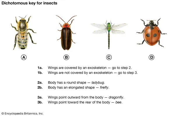
When constructing a key, use observable features, make each pair mutually exclusive, and use one feature at each step.
Organisation of the organism
Levels of organisation
- Cell: the basic unit of a living organism.
- Tissue: a group of similar cells working together.
- Organ: a structure made from different tissues working together.
- Organ system: a group of organs working together.
- Organism: an individual living thing.
New cells are produced by division of existing cells.
Plant and animal cells
| Structure | Function | Plant cell | Animal cell |
|---|---|---|---|
| Cell wall | Supports and strengthens the cell | Cellulose wall present | Absent |
| Cell membrane | Controls movement of substances into and out of the cell | Present | Present |
| Nucleus | Contains genetic material and controls cell activities | Present | Present |
| Cytoplasm | Where many chemical reactions occur | Present | Present |
| Chloroplast | Absorbs light for photosynthesis | Present in photosynthetic cells | Absent |
| Ribosome | Site of protein synthesis | Present | Present |
| Mitochondrion | Site of aerobic respiration | Present | Present |
| Permanent vacuole | Contains cell sap and helps maintain turgor | Large and usually present | No large permanent vacuole |
Identify these structures in labelled diagrams and images. A plant cell also has a cell wall, chloroplasts, and a large permanent vacuole; an animal cell does not.
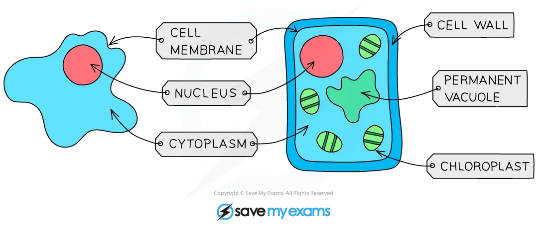
Bacterial cells
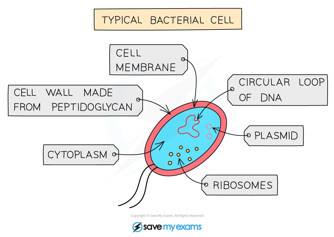
Bacteria are prokaryotes. A bacterial cell has:
- a cell wall for support and protection;
- a cell membrane controlling movement of substances;
- cytoplasm where chemical reactions occur;
- ribosomes for protein synthesis;
- circular DNA containing genetic information; and
- plasmids, which are small circles of DNA separate from the main circular DNA.
Bacterial cells have no nucleus. Identify these structures in diagrams.
Specialised cells
| Cell | Adaptation and function |
|---|---|
| Ciliated cell | Cilia beat to move mucus and trapped particles in airways, or to move an egg cell along an oviduct. |
| Root hair cell | Long projection gives a large surface area for water and mineral-ion uptake; thin wall gives a short path. |
| Palisade mesophyll cell | Near the upper leaf surface and contains many chloroplasts, so it absorbs light efficiently for photosynthesis. |
| Neurone | Long axon carries electrical impulses over a distance; branched ends connect with other cells. |
| Red blood cell | Biconcave shape gives a large surface area; no nucleus leaves room for haemoglobin; flexible shape helps it pass through capillaries; transports oxygen. |
| Gamete | Sperm has a flagellum for movement and many mitochondria for energy. An egg cell is large and contains a food store; both carry genetic material for fertilisation. |
Magnification and measurement
Use: $ [ \text{magnification} = \frac{\text{image size}}{\text{actual size}} ]$
Rearrange when needed:
$$ [ \text{actual size} = \frac{\text{image size}}{\text{magnification}} \qquad \text{image size} = \text{magnification} \times \text{actual size} ] $$
Keep units the same before calculating. For Supplement questions:
- $(1,\mathrm{mm} = 1000,\mu\mathrm{m})$
- $(1,\mu\mathrm{m} = 0.001,\mathrm{mm})$
Worked example
An image is 5 mm long at ($\times 5000$) magnification. Find the actual size.
$[ \text{actual size} = \frac{5,\mathrm{mm}}{5000} = 0.001,\mathrm{mm} ]$
Convert to micrometres:
$[ 0.001,\mathrm{mm} \times 1000 = 1,\mu\mathrm{m} ]$
Answer: 1 μm. Always include the unit.
Typical Sizes in Biology
| Organism/Structure | Approximate Size Range |
|---|---|
| Viruses | 20-300 nm (0.02-0.3 μm) |
| Bacteria | 1000–5000 μm (1-5 mm diameter, typical rod-shaped bacteria like E.coli ~1μm × 2-4μm length) |
| Unicellular organisms | 10–500 μm depending on species (yeast ~7μm, amoeba ~300-600μm) |
| Plant Epidermal Cells | 10-50 μm wide |
| Animal Epithelial Cells | 20-50 μm |
| Red Blood Cells | ~7.5 μm diameter |
| Sperm Cell (HUMAN) | Tail length ~60 μm, head ~5 μm |
| Human Hair Diameter | 50–100 μm depending on individual |
Exam checks
- Use all seven characteristics, not only “living things move”.
- Do not confuse a cell wall with a cell membrane.
- Bacteria have circular DNA and plasmids but no nucleus.
- Name plant and animal groups using observable features.
- In magnification calculations, convert image and actual sizes to the same unit first.