Transport in humans
Circulatory system
The human circulatory system contains:
- vessels to carry blood;
- the heart, a muscular pump; and
- valves to maintain one-way blood flow.
Single and double circulation
In a fish, there is single circulation:
heart → gills → body tissues → heart
Blood passes through the heart once per complete circuit. Pressure falls as blood passes through the gill capillaries.
In a mammal, there is double circulation:
- pulmonary circulation: heart → lungs → heart;
- systemic circulation: heart → body tissues → heart.
Blood passes through the heart twice per complete circuit. The right side pumps blood to the lungs; the left side pumps blood to the body. Double circulation keeps pressure high after blood returns from the lungs and allows efficient oxygen delivery to active tissues.
The heart
Structures and functions
- Atria: upper chambers that receive blood.
- Ventricles: lower chambers that pump blood out of the heart.
- Muscular walls: contract to create pressure.
- Septum: separates the left and right sides and prevents oxygenated and deoxygenated blood mixing.
- Valves: prevent backflow and keep blood moving in one direction.
- Coronary arteries: supply oxygen and nutrients to the heart muscle.
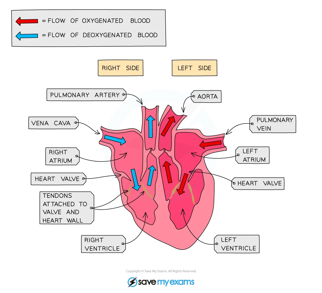
The atrial walls are thinner than the ventricular walls because atria pump blood only into ventricles. The left ventricular wall is thicker than the right ventricular wall because it pumps blood around the whole body at higher pressure; the right ventricle pumps only to the lungs.
Heart function and blood pathway
- The vena cava carries deoxygenated blood from the body to the right atrium.
- The right atrium contracts and the tricuspid valve opens, allowing blood into the right ventricle.
- The right ventricle contracts. The tricuspid valve closes and the pulmonary valve opens; blood leaves through the pulmonary artery.
- In the lungs, blood gains oxygen and loses carbon dioxide.
- Pulmonary veins carry oxygenated blood to the left atrium.
- The left atrium contracts and the bicuspid (mitral) valve opens, allowing blood into the left ventricle.
- The left ventricle contracts. The bicuspid valve closes and the aortic valve opens; blood leaves through the aorta.
- When ventricles relax, the pulmonary and aortic valves close to prevent backflow.
Blood vessels
| Vessel | Structure | Function and pressure |
|---|---|---|
| Artery | Thick muscular and elastic wall; small lumen; no valves | Carries blood away from the heart at high pressure. Elastic recoil maintains flow. |
| Vein | Thinner wall; larger lumen; valves | Carries blood towards the heart at lower pressure. Valves prevent backflow. Skeletal-muscle contractions help move blood. |
| Capillary | Wall one cell thick; very narrow lumen; extensive network | Carries blood close to cells. Short diffusion distance and permeable walls allow exchange of oxygen, nutrients, water, carbon dioxide, and wastes. |
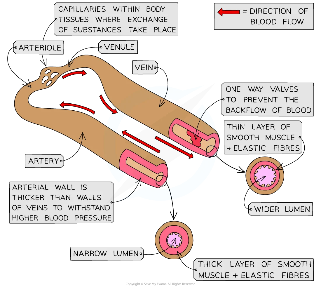
Blood generally becomes deoxygenated in body tissues and oxygenated in the lungs. Exceptions are the pulmonary artery, which carries deoxygenated blood, and pulmonary veins, which carry oxygenated blood.
Main vessels
| Vessel | Route |
|---|---|
| Aorta | Left ventricle to body |
| Vena cava | Body to right atrium |
| Pulmonary artery | Right ventricle to lungs |
| Pulmonary veins | Lungs to left atrium |
| Renal artery | Aorta to kidney |
| Renal vein | Kidney to vena cava |
| Hepatic artery | Aorta to liver |
| Hepatic portal vein | Small intestine to liver; carries absorbed nutrients |
| Hepatic veins | Liver to vena cava |
Blood components
| Component | Structure or appearance | Function |
|---|---|---|
| Red blood cells | Small biconcave cells with no nucleus; flexible and packed with haemoglobin | Haemoglobin binds and transports oxygen. The biconcave shape gives a large surface area and short diffusion distance. |
| White blood cells | Larger, nucleated cells with variable shapes | Defend against pathogens by phagocytosis or antibody production. |
| Platelets | Small cell fragments | Start blood clotting at damaged blood vessels. |
| Plasma | Liquid part of blood, mostly water | Transports dissolved nutrients, hormones, carbon dioxide, urea, ions, and heat. |
In microscope images, red blood cells are numerous, small, regular, and lack nuclei. White blood cells are fewer, larger, and have visible nuclei.
| Cell in an image | Identification clues | Image |
|---|---|---|
| Red blood cell | Numerous, small, regular biconcave cells with no visible nucleus. |  |
| White blood cell | Fewer, larger cell with a visible nucleus and less regular shape. |  |
White blood cells
- Phagocytes engulf and digest pathogens by phagocytosis.
- Lymphocytes produce antibodies. Each antibody has a complementary shape to a specific antigen on a pathogen.
Blood clotting
Clotting prevents excessive blood loss and helps prevent pathogen entry through damaged skin.
- Platelets collect at the damaged area and release clotting factors.
- Soluble fibrinogen is converted to insoluble fibrin.
- Fibrin forms a mesh across the wound.
- The mesh traps blood cells and forms a clot that seals the vessel.
Heart monitoring and exercise
- An ECG records the electrical activity of the heart.
- Pulse rate is the number of heartbeats per minute, measured where an artery is near the skin.
- Valve sounds are caused by valves closing.
During physical activity, muscles respire faster and need more oxygen. The heart rate increases, so more blood and oxygen reach the muscles and carbon dioxide is removed faster. Heart rate usually falls during recovery; recovery can be monitored by measuring pulse rate at regular intervals.
Coronary heart disease
Coronary heart disease occurs when fatty deposits narrow or block coronary arteries. Reduced blood flow deprives heart muscle of oxygen and can cause a heart attack.
Risk factors include:
- a diet high in saturated fat or salt;
- lack of exercise;
- stress;
- smoking;
- genetic factors;
- increasing age; and
- sex-related differences in risk.
A balanced diet and regular exercise reduce risk by helping maintain healthy body mass, blood pressure, and blood lipid levels. Not smoking and managing stress also reduce risk.
Exam checks
- Arteries carry blood away from the heart; veins carry blood towards it.
- The left ventricle has the thickest wall because it pumps around the whole body.
- The septum prevents mixing of oxygenated and deoxygenated blood.
- State both the structure and the function of a capillary.
- For clotting, include fibrinogen → fibrin mesh.
- Do not identify blood vessels only by oxygen content: pulmonary vessels are exceptions.